Monday, January 29, 2018

METABOLIC STABILITY IN VITRO ASSAYS

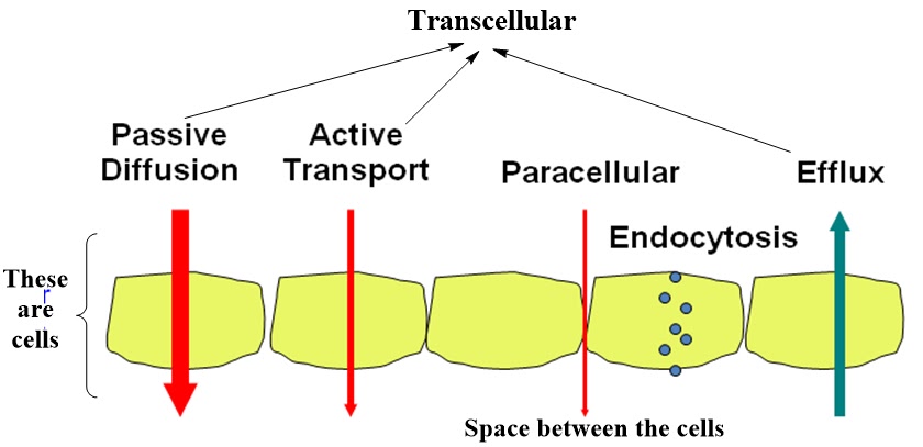
Will the test compound / drug compound remain circulating in plasma within the body?
As mentioned earlier the liver is the major drug metabolizing organ for the large majority of pharmaceutical drugs.  For this reason, the in vitro models used to investigate drug metabolism often focus on hepatocytes or subcellular fractions of the liver such as microsomes, cytosol, S9 or mitochondria where concentrations of particular enzymes are high.

Microsomes contain Phase I oxidative enzymes including CYP enzymes. 

Liver microsomes are subcellular fractions which contain membrane bound drug metabolizing enzymes.  These do not have an intact cell membrane.

The assay uses subcellular fractions of liver, microsomes, to investigate the metabolic fate of compounds. Liver microsomes consist mainly of endoplasmatic reticulum and contain many drug-metabolizing enzymes, including cytochrome P450s (CYPs), flavin  monooxygenases, carboxylesterases, and epoxide hydrolase.

Liver microsomes are available commercially as frozen preparations that are usually prepared in bulk with pooled livers from sacrificed mice, rat or human cadavers (corpse/dead bodies).
Addition of relevant co-factor to the incubation is necessary (NADPH and UDPGA are added.  These are cofactors which are needed to initiate the Phase I metabolic reactions)

The use of species-specific microsomes can be used to enable an understanding of interspecies differences. [for example along with HLM (Human liver microsomes) – MLM (mouse liver microsomes) and RLM (rat liver microsomes) are used]

___________

Most often, metabolic stability of compounds are assessed at a single concentration (typically 10 μM) at t = 0 and at t = 60 min. Stability of compounds are tested in human (other species available) liver microsomes.

___________

S9 fraction is the post-mitochondrial supernatant fraction.  It contains both cytosolic and microsomal enzymes.  This is to identify if cytosolic enzymes are responsible for the formation of a metabolite. 

The advantage of using S9 fraction for in vitro screening is that it contains a wide variety of both phase I and phase II enzymes. 

Addition of relevant co-factor to the incubation is necessary [cofactors such as UDPGA and PAPS to investigate Phase II metabolic pathways.]

Hepatocytes are more representative of the in vivo situation because they contain a cell membrane and do not require additional co-factors.  Hepatocytes contain full complement of enzymes for both Phase I and Phase II metabolism.

The use of species-specific cryopreserved hepatocytes can be used to enable an understanding of interspecies differences in metabolism.

CYP inhibition studies: 

·         CYPs constitute a superfamily of heme enzymes

·         CYPs play a major role in the metabolism of a wide array of xenobiotics including drugs, chemical carcinogens, insecticides, petroleum products, and other environmental pollutants.

·         Although the liver is the primary organ for drug metabolism, extrahepatic tissues such as lung, kidney and intestine, also play an important role for detoxification or biotransformation of xenobiotics. Each tissue has a unique P450 isozyme distribution and regulatory mechanism for cytochrome P450 gene (CYP) expression.


v  In vitro cytochrome P450 inhibition data are useful in designing strategies for investigating clinical Drug-Drug interaction Studies

v  Assessment of the potential of a compound to inhibit a specific cytochrome P450 enzyme is important as co-administration of compounds may result in one or both inhibiting the other’s metabolism. This may affect plasma levels in vivo and potentially lead to adverse drug reactions or toxicity

v  CYP1A2, CYP2C8, CYP2C9, CYP2C19, CYP2D6, CYP3A4 are few important CYP450 enzymes.  Identifying which of these enzymes are responsible for drug metabolism is important.

v  All of these cytochrome P450 inhibition reactions are incubated separately for each isoform.

v  CYP450 3A4 is of paramount importance, because it is the most abundant P450 in the human liver and is known to metabolize the majority of drugs whose biotransformation is known.

METABOLISM and STABILITY OF THE COMPOUNDS/DRUGS


ü  Drug metabolism means the necessary changes in the drug molecules which are essential for the easy excretion from the body. 

ü  Drug metabolism can result in toxication or detoxication - the activation or deactivation of the chemical. While both occur, the major metabolites of most drugs are detoxication products.

ü  Metabolism of the drug is nothing but biotransformation of it.  We already learnt that drug’s are chemically and enzymatically modified to more water soluble compounds (called metabolites).

ü  In general – drug metabolism reactions are divided into two type of reactions →                         Phase I reaction & Phase II reaction


After the drug is absorbed by the GI tract, it is taken up by the part of the bloodstream called the hepatic portal system. Most of the drugs are absorbed into this system except for the lipids which are absorbed into the lymphatic system and then delivered into the blood by the thoracic duct into the superior vena cava. 
The hepatic portal system is designed to take digested foodstuff into the liver where it can be processed, in some cases it is stored before being distributed and it is possible that this may happen to the drug and the drug would be metabolized before reaching the rest of the body. Such drugs that metabolized by the liver are said to have a high hepatic first pass. Hence drugs with a very high hepatic first pass cannot be given orally.
The Blood Brain Barrier:  The capillaries in the CNS are different they have pores which are sealed by the connective tissue and hence only small molecules can cross the blood brain barrier and the substances that can cross over have to be very lipophilic in nature. The blood-brain barrier (BBB) is the protective mechanism of the CNS and is not present everywhere in the brain. This is sometimes useful as it avoids some drugs from crossing the CNS and causing deleterious effects.

Phase I reaction (FIRST PASS METABOLISM) : Here generally oxidation, reduction and hydrolysis type of reactions occur.  Types of enzymes involved in such reactions include cytochrome P450 (CYP) superfamily, flavin-containing mono-oxygenases (FMO), monoamine oxidases, dehydrogenases (for alcohol or aldehyde), reductases, esterases, amidases and epoxide hydrolases.

Such type of metabolism occurs before a drug reaches the systemic circulation.  Typically metabolism occurs in the gut and / or liver before reaching the systemic circulation.

Two thirds of drugs cleared by metabolism are metabolized in part by the cytochrome P450 enzymes with CYP3A4 accounting for almost 50% CYP activity.

One third of the top 200 prescribed drugs which undergo drug metabolism are substrates for metabolic clearance mediated by enzymes other than CYPs. 

Phase II reaction (SECOND PASS METABOLISM) : Here generally addition (or conjugation) of highly polar groups to the drug or drug metabolites occur.   Types of reactions involved include glucuronidation, sulphation, methylation, N-acetylation and glutathione conjugation.


It is not necessary that Phase I reaction occurs first and then Phase II reaction.  If suitable functionalities are present on the drug molecule then Phase II reaction can occur directly.


___________________________


Chemical Stability of the compound:  As discussed earlier, compounds/drugs can be degraded by both chemicals and enzymes in the body. 

Let us first talk about how non-enzymatic degradation process of the compound is measured in a in vitro process.

Test compound is incubated with buffer of interest at different pH (commonly pH2, pH6 and pH10).  Some buffers which are also investigated include simulated intestinal fluid (SIF) and simulated gastric fluid (SGF). 

At different time intervals the concentration of test compound is quantified by LC-MS. 

Plasma Stability of the compound: 

In addition to hepatic metabolism, compounds are also subjected to degradation/ modification by enzymes in plasma.  Instability of compound/drug in plasma generally show poor in vivo efficacy.  Investigation of plasma stability should be performed early in the discovery process in order to assess potential degradation and/or protein binding issues.

In general, esters, amides, lactones, lactams, carbamides, sulphonamides, and peptic mimetics tend to more susceptible to hydrolysis in plasma.

___________________________

A solution of test compound in plasma is prepared and incubated for a predetermined time period. Aliquots are removed at pre-defined time points and analyzed by LC/MS/MS. The peak area for the parent compound is compared to the time zero sample in order to assess the amount of compound still available.
___________________________

Sunday, January 21, 2018

ABSORPTION: PASSIVE AND ACTIVE TRANSPORT (PERMEABILITY) -2


Parallel Artificial Membrane Permeation Assay (PAMPA):   

As the name suggests, this is a cell-free assay to predict passive, transcellular permeability of the drugs in early drug discovery.  These artificial membranes have no active transport systems and  no metabolizing enzymes, moreover this assay would not provide us the information regarding actively transported molecules.  This gives purely passive diffusion of unchanged compound.


The ability of this assay to evaluate permeability over a large pH range is valuable for an early understanding how new oral compounds might be absorbed across the entire gastrointestinal tract.


·         The artificial membrane is first built-up by pipetting a solution of lipids on a supporting filter material in micro titer plates ( say 96-well plates)

·         There should also be a control well where lipid layer is absent.

·         Test compounds are added to the donor compartment.

·         Permeation occurs through the artificial membrane into the acceptor compartment

·         Now compound permeated to the acceptor compartment is measured photometrically or by LCMS.


In few protocols, 96-well microtitre plate and a 96-well filter plate is sandwiched such that each composite well is divided into two chambers: donor at the bottom and acceptor at the top.

The gastrointestinal tract (GT) has a pH range from pH 1 – 8. The pH of the blood is constant at pH 7.4; therefore it is possible for a pH gradient to exist between the GT and the plasma that can affect the transport of ionizable molecules. In an effort to mimic this pH gradient in vitro, alternative assays with pH 7.4 for the acceptor compartment and pH values 5.0, 6.2, and 7.4 in the donor compartment are used.


Permeability values are reported in (10-6 cm/s)


Compounds which have a

PAMPA: Papp < 10 x 10-6cm/s are classified as low permeability and 
               Papp > 10 x 10-6cm/s are classified as high permeability
___________________________

Caco-2: 

The Caco-2 cell line is derived from a human colon (large intestine) carcinoma.

The cells have characteristics that resemble intestinal epithelial cells such as the formation of a polarized monolayer, well-defined brush border on the apical surface and intercellular junctions.

Caco-2 cells express tight junctions, microvilli, and a number of enzymes and transporters that are characteristic of such enterocytes lining the small intestine.


     ·         Caco-2 human colon adenocarcinoma cells are grown to confluence (to form a monolayer covering the surface of the filter). 

·         Test compounds are added to apical side of the compartment.

·         Permeation occurs through the top side of the monolayer,

·         Now compound permeated to the basolateral compartment is measured photometrically or by LCMS.

·         Apparent permeability coefficient (Papp in cm/s) is determined. 
Rule of thumb numbers for Caco-2 permeability 

Low Caco-2 permeability (<5 x 10⁻⁶ cm/s) ; high Caco-2 permeability (>20 x 10-6 cm/s)

Caco-2 assay allows us to assess transport of the drug in both directions i.e. apical to basolateral (A-B) and basolateral to apical (B-A)) across the cell monolayer. (known as bi-directional Caco-2 permeability assay)

Thus enabling us to determine an efflux ratio which provides an indicator as to whether a compound undergoes active efflux.

The result is typically reported as an efflux ratio i.e. Papp(B-A)/Papp(A-B). If the efflux ratio is greater than two (Papp(B-A)/Papp(A-B) > 2) then this indicates drug efflux is occurring.

PAMPA is a pre-screening tool in early drug discovery.  Molecules/compounds which behaved well in PAMPA assay (permeated well) are further test with Caco-2 model.

PAMPA allows high throughput screening of molecules (compared to Caco-2)

Caco-2 model is more descriptive – as it gives passive as well as active transport. 


Madin-Darby Canine Kidney (MDCK) cells are also used in place of Caco-2 cells for permeability assays.  These cells can also be grown as a monolayer with tight junctions.  However, MDCK cells are from canine source (dog or dog-like mammals).  Moreover, these are kidney-derived, not intestine-derived as Caco-2 cells.  Therefore, given a choice Caco-2 is more desirable than MDCK.

___________________________

Ø  MDR1-MDCK cells are common choice for evaluating P-gp substrates and inhibitors.  Cell lines are transfected with transporters of interest.  MDR1 is the gene encoding for the efflux protein, P-glycoprotein (P-gp)

Ø  P-gp is one of the mosot well-recognized efflux transporters in many tissues including the brain, kidney and intestine.

Ø  Therefore, MDR1-MDCK has been found to be useful predictor of blood brain barrier permeability.


MDR1-MDCK Papp (apical to basolateral) → gives an indication of the extent of permeation across cells which express P-gp (e.g. gastrointestinal tract and blood brain barrier)

Efflux ratio → gives the extent of drug efflux by P-gp


Even Caco-2 study will give us Efflux ratio [by performing bidirectional assay].  Exactly same as above, by assessing transport in both directions (apical to basolateral (A-B) and basolateral to apical (B-A)) across the cell monolayer enables an efflux ratio to be determined.  Thus, we can know if the compound undergoes active efflux.

Add a P-gp inhibitor to suppress the efflux – the result obtained confirms the role of P-gp in the efflux

Add a known P-gp substrate A – then add the test compound B → If the test compound is P-gp inhibitor then substrate A becomes permeable.  Thus test compound B’s P-gp inhibitory effect can be evaluated.

NOTE
Caco-2 experiments also do not predict paracellular absorption well because the paracellular route is tighter in Caco-2 cells than that in the small intestine in vivo. While the average pore radius of the tight junctions in the human small intestine is around 8–13Å, the corresponding radius in Caco-2 cells is about 4Å. When the paracellular pathway is narrower, the intrinsic permeability will be lower than in the in vivo situation.

___________________________

ABSORPTION: PASSIVE AND ACTIVE TRANSPORT (PERMEABILITY) -1


(1)   During the travel of Oral Drug from mouth to intestine, through-out its journey drug has a chance to get absorbed in to blood and also faces metabolism problem.

(2)   Most of the absorption of the drug takes place in the small intestine. Since the surface area of the stomach (and all other parts starting from mouth to stomach) is much smaller than that of the intestine.  The amount of time that the drugs spend in the stomach (and all other parts starting from mouth to stomach) is less and also the surface area of the stomach is small.


·         For a drug to be absorbed in the intestine some portion of it needs to be dissolved in the intestinal juices which are aqueous.  Even if drug is lipophilic the above said dissolution of drug in the intestinal juices is necessary.  

·         Therefore, in case of partly soluble drugs in water, whatever amount is soluble first gets absorbed and then an equivalent amount of un-dissolved portion of drug becomes soluble which is now ready to get absorbed.  Thus complete absorption will take place.

·         There are bile salts present in the intestine which will aid in solvation of the drug and their resultant absorption. Drugs that are amphipathic have no problem in getting absorbed. There are some drugs that are completely insoluble in water such drugs float as globules in the intestine but the bile salts will emulsify these into small enough particles such that absorption can take place.

·         Some drugs can be absorbed into the system by active transport.

____________________________

Once the drug is in the body – it has to cross several barriers to reach the site of action.

Barriers like - Gut wall (passage across GI tract wall), cross lipid barriers / cell wall, blood brain barrier etc.  Therefore, bioavailability of the drug depends on all these parameters.

Let us first understand how the compounds permeate through these barriers or membranes.  What are the different permeation mechanisms? 

·         Drug permeation involves several processes of drug transport across the cell membranes.
·         We know that drugs are most often administered away from their site of action
·         Therefore, drug has to permeate from one compartment to another crossing different barriers and then cell membranes.
·         Drug in the GI tract has to pass through the intestinal membrane.  We are going to talk different assays to predict intestinal permeability.  

·         Drug crosses the membranes and gets into the cell by different mechanisms,
i.                    Passive transport (passive permeability) –MAJOR ABSORPTION PATHWAY
ii.                  Active transport (active permeability) – Carrier mediated transport
Drugs are foreign molecules to the cells.  There are specialized carriers which expel these foreign molecules as they enter the cells.  These are called reverse transporters (efflux transporters).


Below is one more cartoon diagram of the same thing presented above.


TWO most common in vitro assays to obtain permeability data:
Parallel Artificial Membrane Permeation Assay (PAMPA) and Caco-2